
|
Boletín de la Sociedad Geológica Mexicana Volumen 74, núm. 3, A200722, 2022 http://dx.doi.org/10.18268/BSGM2022v74n3a200722
|
 |
Subducted iron and glassy spherules in the upper mantle?
¿Esférulas de hierro y vítreas subducidas en el manto superior?
José M. González-Jiménez1,2,*, Ivanina Sergeeva3, Thomas N. Kerestedjian3, Fernando Gervilla1,2
1 Instituto Andaluz de Ciencias de la Tierra, Consejo Superior de Investigaciones Científicas (CSIC)-Universidad de Granada, Avda. de las Palmeras 4, 18100, Armilla, Granada, Spain.
2 Departamento de Mineralogía y Petrología, Facultad de Ciencias, Universidad de Granada, Avda. Fuentenueva s/n, 18002, Granada, Spain.
3 Geological Institute, Bulgarian Academy of Sciences, 24 Georgi Bonchev Str., 1113, Sofia, Bulgaria.
* Corresponding author: (J.M. González- Jiménez) This email address is being protected from spambots. You need JavaScript enabled to view it.
How to cite this article:
González-Jiménez, J.M., Sergeeva, I., Kerestedjian, T.N., Gervilla, F., 2022, Subducted iron and glassy spherules in the upper mantle?: Boletín de la Sociedad Geológica Mexicana, 74 (3), A200722. http://dx.doi.org/10.18268/BSGM2022v74n3a200722
Manuscript received: June 26, 2022; Corrected manuscript received: July 19, 2022; Manuscript accepted: July 24, 2022.
ABSTRACT
Spherules are documented in ophiolitic mantle rocks such as peridotites and associated chromitites. They consist of: (1) native iron having variable amounts of Ni with/without inclusions of silicate glass or oxides (wüstite), (2) dendritic intergrowth of oxides (magnetite, wüstite and hematite) with/without silicate glass and, (3) silicate glass. Consensually, they are interpreted as indigenous to chromitites and related with high-temperature processes operating in the Earth’s upper mantle. However, their similarity with terrestrial and extraterrestrial spherules found in other settings of the geological record is remarkable. We raise the question on such indigenous origin, relating them to volcanic and cosmic material recycled back to the mantle wedge where chromitites form during subduction.
Keywords: spherules, iron, glass, upper mantle, chromitite, subduction.
RESUMEN
Rocas del manto superior ofiolitico tales como peridotitas y cromititas contienen esferas. Estas consisten de: (1) hierro native con cantidades variables de Ni con y sin inclusiones de vidrio silicatdo y óxidos (wüstite), (2) intercrecimientos dendríticos de óxidos (magnetita, wüstita y hematites) con o sin vidrio silicatado y, (2) vidrio silicatado. Unísonamente, estas esferulas se interpretan como indígenas a las cromititas y relacionadas con procesos de alta temperatura que tienen lugar en el manto superior terrestre. Sin embargo, su parecido con aquellas esferas de origen terrestre y extraterrestre descritas en otros contextos geológicos es reseñable. En este trabajo cuestionamos el origen autóctono de las esferas en las rocas mantélicas, interpretándolas como material de origen volcánico y cósmico que ha sido reciclado atraves de la cuña de manto superior donde ser forman las cromititas durante los procesos de subducción.
Palabras clave: esferas, metal, vidrio, manto superior, cromitita, subducción.
- Introduction
Millimetric to (sub)-microscopic metallic-silicate-oxide spherules of both extraterrestrial and terrestrial origins are present in the fossil and current geological record. The extraterrestrial ones are part of the huge number of micrometeorites and cosmic dust that rains on the Earth annually resulting from the fragmentation of asteroids and released from comets (Peucker-Ehrenbrink, 2001). Such extraterrestrial material enters the atmosphere and partially melts and oxidizes to form tiny (<2 mm) spheroidal bodies consisting of iron or iron-nickel oxides and/or alloys (I-type), Fe oxide dendrites within silicate glass (G-type) or mainly silicate (S-type; with the V subtype made up only of silicate glass) (Genge et al., 2008). On Earth, metallic-silicate-glass spherules may form by: (1) high-temperature processes but related with volcanism (i.e., cloud-to-ground lightning strikes or eruptions of low-viscosity magmas; Genaerau et al., 2015; Nyström et al., 2016), (2) meteoritic impact (i.e., crystallization from the splash ejecta or condensation from rock-vapor plumes; Velázquez et al., 2021), (3) biogenic hydrothermal activity (Agarwal et al., 2021), and (3) diagenesis (Suk et al., 1990). Likewise, un-natural anthropogenetic industrial processes may also manufacture spherules (Niyogi et al., 2011; McCloy, 2019).
Metallic-oxide-silicate spherules are also found in mantle rocks (peridotites and chromitites) from a few ophiolites (Bai et al., 2004; Robinson et al., 2004; Xu et al., 2009; Xu et al., 2015; Yang et al. 2015; Griffin et al., 2016), although its origin is highly controversial. They are interpreted to from within mantle itself by melt immiscibility under the highly reducing conditions (e.g., Griffin et al., 2016; Xiong et al., 2017) or to be solid products of molten droplets originated by lightning strikes on the mantle rocks when exposed to Earth’s surface (Ballhaus et al., 2017). Here we provide the first-ever thoughtful comparative study of metallic-oxide-silicate spherules from ophiolitic upper mantle rocks with others present in the geological record, raising the possibility that they could correspond to recycled airborne spherules of cosmic and volcanic origin that originally landed on the deep-sea floor were recycled back to the mantle during subduction. Our discovery offers an innovative alternative to foster the current debate of exotic minerals in ophiolitic rocks while unravelling geodynamic processes that operate in subduction-zone settings.
- Analytical methods
2.1. MINERAL CONCENTRATES
A small chip from each of the four samples analyzed in this study (GK1C-1 and GK3A-1 from Golyamo Kamenyane and J-410m and Y-409m from Yakovitsa) was pre-grounded in agate mortar, washed with ethanol and then dried at room temperature for few minutes (until complete evaporation of the ethanol). Dry material was then divided into sub fractions by handheld magnet, pre-insulated from direct contact with the material to avoid contamination, and purified several times. In order to avoid potential contamination, the manipulations of washing with ethanol were done in glass Petri dish and the acid treatment in PTFE containers (100 ml tanks). This way, two fractions were separated – non-magnetic, silicate one, and magnetic chromite one. As a precaution, the material was inspected under a binocular microscope at each step of the separation process and no spherules were detected at this stage. Each fraction was subsequently separated by granulometry using 200 mesh disposable sieves while the fraction 70-100 µm was again checked for spherules, although they were not identified. The chromite fractions were further treated with a mixture of HF and HCl acid, heated to 160° C. The insoluble residue was then washed with deionized water several times and dried at room temperature revealing the presence of spherules when recognized under binocular microscope. Once recognized, the spherules were hand-picked and sticked on a wire of carbon tape on a SEM pin stub holder to be imaged under scanning electron microscopy. Subsequent to this preliminary analysis of the spherule surface, then they were imbibed in epoxy resin and polished. Considering the small size and their softness, the largest iron spherule identified from the Yakovitsa chromitite was the only to survive the polishing process.
2.2. FIELD EMISSION SCANNING ELECTRON MICROSCOPY (FE-SEM)
Spherules were preliminarily characterized and imaged using a Leo Gemini Field Emission Scanning Electron Microscope (FE-SEM) at the Centro de Instrumentación Científica (CIC) of the Universidad de Granada, Spain. The instrument was equipped with an Energy Dispersive Spectra (EDS) detector. Accelerating voltage was 20 kV and beam current optimized for an adequate number of counts for each EDS analysis. The unpolished spherules were firstly inspected on the secondary electron mode (SE) in an effort to better define their morphological characteristics and chemistry by EDX, where once polished they were checked again under FESEM but using the back-scattered electron mode (BSE), although only one of the spherules survived (i.e., Spherule 1 from Yakovitsa shown in Figure 2 of the main text).
2.3. MICRO-RAMAN
The micro-Raman spectra of silicate and oxide component of the spherules were obtained by means of JASCO NRS-5100 micro-Raman spectrometer, equipped with an Olympus microscope with x5, x20, and x100 objectives belonging to the CIC of University of Granada, Spain. The 532 nm laser excitation was used as an excitation source with a spectral resolution of 2.08 cm-1. The minimum lateral resolution was about 1 μm (with the 100× objective). The spectrometer was calibrated using the 520.6 cm-1 Raman peak of silicon before each experimental measurement. The spectra were collected using the 100× objective with repeated acquisition: 2 acquisitions for 100 s. Obtained Raman spectra were compared with those of the RRUFF database.
2.4. ELECTRON MICROPROBE ANALYSIS (EPMA)
The major element composition of magnetite was determined on the polished surface for the Spherule 1 using a Cameca SX-100 microprobe in CIC of University of Grana, Spain.
EPMA data were obtained using an excitation voltage of 20 kVand a beamcurrent of 20 nA, with
a beam 2–3 μm in diameter. Monitored spectral lines were MgKα, FeKa, AlKα, CrKα, SiKα, TiKα, MnKα, NiKα, CoKα, ZnKα and VKα. Standards used were MgO, Fe2O3, Al2O3, Cr2O3, SiO2, CoO, ZnO, TiO2, MnTiO3, NiO and Pb5(VO4)3Cl.
2.5. FOCUSED ION BEAM (FIB) HIGH-RESOLUTION TRANSMISSION ELECTRON MICROSCOPY (HRTEM)
A thin-foil sample was prepared and extracted from the polished Spherule-1 from the Yakovitsa chromitite by using a Focused Ion Beam Scanning Electron Microscope (FIB-SEM) in the Laboratorio de Microscopías Avanzadas (LMA) at the Instituto de Nanociencia de Aragón (INA) – University of Zaragoza, Spain. The TEM thin foil preparation was performed using a Dual Beam FEI Thermo-Fisher Scientific, model Helios 650. The selected region of interest was first covered by a thin strip (~300 nm) of C by focused electron beam induced deposition (FEBID) and subsequently with a second strip (~1 µm) of Pt. These strips act as protection during the milling, polishing, and extraction process of the thin foils. The bulk material was first removed on both sides of the lamella by a rough Ga+ ion milling with a 30 kV current at 2.5 nA and the subsequent polishing with a 30 kV current at 0.23 nA. The final polishing step was performed on the sample’s inclusions until the electron transparency was achieved. This was completed by subsequently milling the thin foil with a 5 kV current at 68 pA. The electron transparency was monitored by an Everhart-Thornley SE detector and using a 5 kV electron beam. After achieving the electron transparency, the thin foil was rapidly polished using a low energy 5 kV current at 10 pA to reduce the amorphization until a final thin foil thickness of ~90 nm was attained. Subsequently, the thin foil was undercut with a 30 kV at 2.5 nA current, lifted out, and transferred from the sample to a TEM grid using an OmniProbe nano-manipulator with a tungsten tip. To weld the thin foil to the tungsten tip and the TEM grid, an ion-beam assisted Pt deposition was performed.
A FEI Titan G2 transmission electron microscope (TEM) equipped with Field Emission gun XFEG was used to analyze the thin-foil at the Centro de Instrumentation Científica of the University of Granada, Spain. The FEI Titan G2 microscope is equipped with 4 energy dispersive analyses of X-rays (EDX) detectors (FEI microanalysis Super X) and a high-angle annular dark-field detector (HAADF). This FEI Titan G2 microscope also includes a spherical aberration corrector for the objective lens. Selected mineral areas of interest sampled within the thin-foil were imaged by the FEI Titan G2 microscope using a combination of high-angle annular dark-field (HAADF) to obtain Z high contrast images, and High-Resolution Transmission Electron (HRTEM) images to characterize the texture of the grains and to properly define the ordering of the mineral aggregates. All these images were treated using the Digital Micrograph® software in its Version 1.71.38 while maps were processed with the VELOX® software package. The FEI Titan G2 was running at 300 kV working conditions while HRTEM images were acquired using Gatan CCD Camera. Elemental maps images were also processed using the VELOX® software.
- Results
Two iron-rich oxide and two silicate glass spherules were recovered from two chromitites bodies hosted in the upper mantle section the Golyamo Kamenyane and Yakovitsa ultramafic massifs, in southern Bulgaria (Figure 1). These rocks belong to the Rhodopean Ophiolite Association (Kolcheva et al., 2000), which correspond to a lithospheric section formed a mid-oceanic ridge that evolved to an island arc/back-arc setting in a supra-subduction zone (Colás et al., 2013).
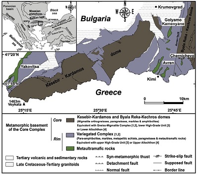 |
| Figure 1. Simplified geological map of the Eastern Rhodope (adapted from Bonev, 2006) showing the localization of the ultramafic massifs hosting the chromitite bodies investigated in this work. Equivalence keys: 1 (Kozhoukharov et al., 1988), 2 (Haydoutov et al., 2001), 3 (Bonev, 2006), and 4 (Janák et al., 2011). Legends are in all cases inset in the figures. |
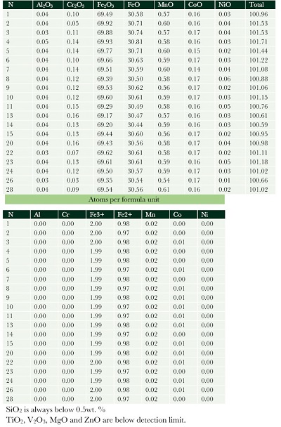
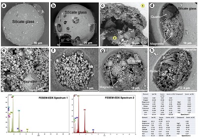
The largest iron-rich oxide spherule (~75-175 µm) was found in the Yakovitsa chromitite (Figure 2a-2l). It consists of an interlocking polygonal structure made up of micro-crystallites of magnetite with junctions, which are filled by a finely-grained symplectite of magnetite and amporphous Si glass with trace amounts of P (Figure 2a-2l). Single-spot analyses obtained by EMPA show relatively homogenous composition of magnetite across the whole sphere with up to 93.47 wt.% FeOt, minor Mn (0.54-0.61 wt%), CoO (0.14-0.17 wt%), Cr2O3 (0.03-0.16 wt%), Al2O3 (0.03–0.05 wt%), NiO (0.01-0.06 wt%), SiO2 (<0.5 wt%), and no TiO2, MgO or CaO (Table 1). The smaller iron-rich oxide spherule identified in Golyamo Kamenyane (Figures 3a-3c) consists of dendritic magnetite (Fe2O3) microcrystals within a wüstite (FeO) groundmass as determined by micro-RAMAN and FESEM-EDX analysis collected from the spherule surface (Figures 3d-3f). The discrete crystals of magnetite are small (<5 µm) faceted-equant grains with cubic arrangement perpendicular to (111) crystal faces (Figure 3b), whereas rosaries of sub-micrometric spherules of magnetite occur widespread in the wüstite groundmass (Figure 3c).
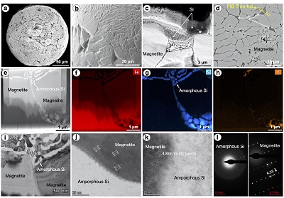 |
| Figure 2. Microphotographs of the iron spherule recovered from the Yakovitsa chromitite. (a-c) and (d) are images of the microtexture of the spherule collected by FE-SEM previously and after polishing, respectively. (e-h) High-angle annular dark-field scanning transmission electron microscopy (HAADF-STEM) image and corresponding TEM-EDS elemental map of the FIB thin foil (see location in (d)). (i-k) High-resolution (HR)-TEM and high-magnification HRTEM images of the anatomy of the contact between magnetite microplates and symplectite amorphous silica. (l) Selected area electron diffraction (SAED) image showing the amorphous nature of the silica sympletitice and crystallinity of magnetite. |
| Table 1. Chemical composition of magnetite. |
 |
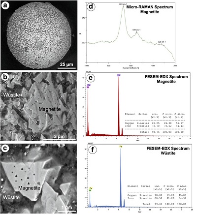 |
| Figure 3. Microphotographs of the iron spherule recovered from the Golyamo Kamenyane chromitite. (a-b) Secondary electron (SE) FE-SEM images of the unpolished spherule. (d-f) Representative spectrums of magnetite and wüstite collected micro-RAMAN and FESEM. |
The glassy spherules are both colorless with a smooth surface (Figures 4a and 4b) relatively homogenous in composition dominated by SiO2 (~41 wt.%), CaO (31-34 wt-%) and Al2O3 (14-15 wt%), and lesser amounts of FeOt (~7 wt.%), MgO (2-4 wt.%) as well as Ti and K < 1.5 wt.% (see EDX inset in Figure 4). These two spherules were partly smashed during the mounting process, reveling internal continuity of the vitreous structure with the typical conchoidal fractures of glassy materials (Figures 4c and 4d). The spherule recovered from the Golyamo Kamenyane chromitite displays shrinkage features such as bubble-like vesicles now filled with a suite of secondary minerals including magnetite, quartz, rutile or mixtures of them (Figure 4b). Under high magnification magnetite exhibit rounded to octahedral-habit, whereas quartz is prismatic and rutile is needle-like to acicular crystals grown perpendicular to the surface of the vesicle.
 |
| Figure 4. Microphotographs collected by FESEM in SE mode of the glassy spherules recovered from the Yakovitsa (a) and Golyamo Kamenenyane (b-h) chromitites. Representative FESEM-EDX spectrum of the glass are inset in the figure. |
- Discussion and concluding remarks
4.1. IDENTIFYING SPHERULES IN MANTLE-DERIVED ROCKS
As noted above, metallic-silicate-oxide spherules found in natural rocks may have both natural and anthropogenetic origins. Some textural (magnetite, rutile and quartz filling voids in the glassy spherule) and chemical features (very low Mn in the iron-rich spherule) of the spherules from the Bulgarian chromitites clearly rules out contamination by man-made spherules (Sulovsky, 2002; Zhang et al., 2014; Shetye et al., 2019) during mineral separation processes.
Spherules having structures and textures similar to ours - dendrite magnetite with interstitial silicate glass or wüstite groundmass and silicate glass only; Figures 2-4 - have not been reported yet from ophiolitic mantle-derived rocks. Most spherules documented in peridotites and associated chromitites from ophiolites (i.e., Tibet, Ray-Iz and Oman) consist of Ni-free native iron (α-Fe) with cortex of dendritic wüstite, both having very small quantities (<<1 wt.%) of Mn, Ti and Si (Bai et al., 2004; Robinson et al., 2004; Xu et al., 2015; Griffin et al., 2016). Some spherules documented from Tibetan chromitites exhibit an additional thin layer of hematite separating the iron core and the wüstite rim (Xu et al., 2015), whereas some others from Ray-Iz have iron core with drop-like inclusions wüstite, Fe–Ti alloys and/or silicate glass with up to 39 wt.% TiO2, 19 wt.% SiO2, 4 wt.% Al2O3, 22 wt.% FeO, 8 wt.% MnO, 6 wt.% Ca and 1 wt.% K2O (Yang et al., 2015). A subset of the spherules identified in-situ in chromites from Tibet consist of Ni-Fe (formula Ni7.4 Fe2.6; Xu et al., 2009) and host inclusions of pentlandite, periclase and magnesium silicates.
These spherules documented in mantle-derived ophiolitic rocks were related to fluids/melts of very high temperature (>1400-1500 ºC) and low fO2 (well below IW buffer) operating in the lower mantle or the Mantle Transition Zone (i.e., MTZ > 610 km; Xu et al., 2015; Griffin et al., 2016). This interpretation was based on two assumptions: (1) the assemblage iron + wüstite was also found in diamonds of deep-seated peridotites and kimberlites (e.g., Xu et al., 2015), and (2) metallic-silicate-oxide spherules belong to the Super-reducing (SuR) mineral assemblage (e.g., moissanite, carbides, nitrides and silicides) that is usually found together with minerals that typically form at UHP (e.g., diamond and coesite) in these mantle rocks. Pioneering works suggested a common origin of the SuR and UHP during chromitite crystallization from high-Cr melts within the lower part of the upper mantle or MTZ (Yang et al., 2007; Zhou et al., 2014; Zhang et al., 2016) or metamorphism of recycled chromitites at these high depths (McGowan et al., 2015; Griffin et al., 2016). However, subsequent works (e.g., Xiong et al., 2017) suggested that SuR minerals could be incorporated into exhuming peridotites (and associated chromitites) that already had UHP by metasomatic slab-derived CH4 ± H2 fluids, or mantle melts that were supplied with such component. Moreover, all spherules identified in these mantle-derived rocks, except the Ni-Fe bearing isolated inclusions in chromite from Tibetan chromitites, were found in mineral separates, therefore do not provide the necessary direct connection between the spherules and the other minerals of the SuR-UHP to confirm their true mantle origin. In addition, subsequent works showed that SuR-UHP minerals might also form during low-pressure partial hydration of oceanic lithosphere (Farré-de-Pablo et al., 2019; Pujol-Solà et al., 2018). Thus, a need for the inquiry on the generalized deep mantle-related origin of for the alleged SuR-UHP assemblage has arisen.
The spherical morphologies along with the droplet-like shape of wüstite and/or silicate glass within iron cores reported in spherules from Ray-Iz are consistent with surface tension of unmixing metallic, oxide and silicate melts at high-temperature. However, such mechanism itself cannot satisfactorily explain the presence of silicate glass found as both inclusions within native iron and interstitial to dendritic magnetite and/or wüstite. Rather, silicate glass suggests melts quickly frozen (quenched) as these are non-equilibrium materials that form due to fast kinetics preventing complete crystallization after melting with extremely high cooling rates (i.e., in the order of 107 ºC/min for hyper-quenched glasses; McCloy, 2019). Therefore, it is unlikely that those spherules containing glasses were originated in-situ within the Earth’s mantle. In fact, silicate glasses in mantle rocks can only be attributed to trapped interstitial melts that have quenched during fast volcanic eruptions (O’Reilly and Griffin, 2012). Interestingly, the glass in these spherules from Ray-Iz has higher Ti than glasses in analogous volcanic and cosmic spherules. Its chemistry is, however, similar to that of silicate-titanium-iron spherules reported from the Colônia impact crater in Brazil (Veláquez et al., 2021). However, these spherules lack of sculpturing or impact marks (microcraters, pits with radial cracks and star-shaped pit) as those of impact-related origin.
The patterns of iron nuclei and oxide cortex or dendritic magnetite in wüstite groundmass documented in the spherules from Tibet and Ray-Iz and Golyamo Kamenyane ophiolites are very similar to those reported in I-type cosmic spherules (Genge et al., 2017). In contrast, the microtexture of dendritic magnetite within Si-bearing glass of the Yakovitsa spherule has been identified in volcanic (e.g., Ignimbrites of Southern Primorye in Rusia; Grebennikov, 2011) and G-type cosmic spherules (Folco and Cordier, 2015). A common feature of all spherules found in ophiolitic mantle rock is that both magnetite and wüstite have Ni contents below 1.0 wt.%. Low Ni in oxides is a common feature of cosmic spherules where Ni was separated from the silicate-oxide melts towards metallic beads due to siderophile attraction display (Genge et al., 2017). In fact, a population of spherules reported from Tibetan chromitites still contain Ni-Fe beads or are solely Ni-Fe spherules, similarly to the vast majority of I-type cosmic spherules reported in the geological record (Folco and Cordier, 2015; Genge et al., 2017). Nevertheless, cosmogenic nuclide and noble gas analyses are not available for spherules associated with mantle peridotite and chromitites to confirm their extraterrestrial or terrestrial origin. This is clearly a green field for future research.
Silicate glass spherules from our chromitites exhibit some common features with silicate-dominant volcanic and V-type cosmic spherules, including: (1) spherical morphology, with smooth external surface and internal conchoidal fractures as is typical for quenched silicate-droplet glass, (2) empty vesicles (now filled with secondary minerals) that may have been produced by degasification or dissolution of pre-existing metal beads. However, their markedly higher Al and Ca but lower Mg distinguish them from cosmic ones. Overall, the concentration of all the analyzed elements overlaps that of terrestrial basic basaltic glasses (McCloy, 2019; Cicconi and Neuville, 2019), suggesting their possible volcanic-related origin.
4.2. RECYCLING SPHERULES IN THE UPPER MANTLE?
Shi et al. (2011) suggested an extraterrestrial origin for a suite of iron spherules they recognized in jadeitites from the Myanmar ophiolite. In these spherules with Fe isotopic composition overlapping I-type cosmic spherules, core of native iron is also overgrown by a rim made up of dendritic crystal of wüstite. This discovery shows that cosmic spherules could remain stable under low-temperature and high-pressure conditions prevailing upon subduction, and therefore might be found in rocks related to slab-derive sediments in subduction zones. Interestingly, some spherules from Ray-Iz exhibit an identical pattern, raising the possibility that cosmic spherules (and by extension volcanic ones) now found in peridotites and chromitites from ophiolites could represent part of sediments that were incorporated into the upper mantle via subduction. The recycling of crustal material in the mantle and return pathways to the Earth’s surface has been already tracked by xenocrystal zircons inherited from continental sources in supra-subduction zone ophiolitic and juvenile intra-oceanic volcanic arc rocks (Proenza et al., 2018). In this scenario, it is expected that the spherules had experienced a series of events since their deposition in the deep sea including: modification during fly, alteration during diagenesis and transport with the oceanic slab into the subduction zone, high P/T metasomatism/metamorphism, heating by carrying fluids and melts involved in mantle metasomatism and chromitite formation, and final exhumation. However, all the spherules identified in ophiolitic mantle rocks are well-preserved as their shapes and chemical composition were not obviously altered. Then, how can these spherules could be transported and recycled into the shallow oceanic mantle without affection?
Subducted slabs can transport significant volumes of sediments to depths greater than 50 km (Scholl and von Huene, 2009) and these, in turn, may be transferred to the mantle wedge by (1) fluids/melts channelized in melt conduits or fractures (González-Jiménez et al., 2017), and/or (2) detachment and buoyancy-driven diapiric up flow of plumes of slab-derived sediments (Gerya and Yuen, 2003). The first mechanism links the transfer of slab-derived spherules to the introduction of silica- and alkali-rich fluids/melts due to the dehydration/ anatexis of subducted sediments through fractures in mantle peridotites; it could be linked to mechanism similar to those transferring spherules to jadeitites as observed in the Myanmar ophiolite. The second mechanism could be related with the cold plumes –partly molten crustal (including sediments, continental crust and basalts) and mantle rocks mixed on length scales from a few tens of meters to hundreds of kilometers – contributing to mantle metasomatism and chromitite formation (González-Jiménez et al., 2017; Proenza et al., 2018). In these two scenarios, the equilibration of spherules with surrounding rocks and melts might be inhibited by their rapid transfer through fractures or by porous flow in small melt pockets from the melting source to a new site in, or their inclusion in shielding minerals like pyroxene, olivine or during melt transport. The incorporation of such type of inherited xenocrystic pyroxenes and olivine have been largely reported in chromitites and melt pockets of peridotites from ophiolites (e.g., Borisova et al., 2012; Griffin et al., 2016).
Contributions of authors
JMGJ and IS designed the experiment and carried out the analytical work. JMGJ wrote the manuscript which was revised and intellectually improved by the input of TNK and FG.
Financing
Funding for analytical work was provided by the RNM-131 Group of the Junta de Andalucía “Grupo de Investigación Mineralogía Petrología y Yacimientos Minerales (GIMPY)”.
Acknowledgments
The authors acknowledge to Bendicion Garcia-Furnes, Miguel Ángel Hidalgo-Laguna, Maria del Mar Abad and Cecilia de la Prada their assitance for analytical work at the Centro de Instrumentación Científica of the University of Granada.
Conflicts of interest
The authors declare no conflict of interest.
References
Agarwal, D.K., Palayil, J.K., 2021, Recovery of Hydrothermal Wustite-Magnetite Spherules from the Central Indian Ridge, Indian Ocean: Research Square. https://doi.org/10.21203/rs.3.rs-948343/v1
Bai, W., Yang, J., Fang, Q., Yan, B., Zhang, Z., Ren, Y., Shi, N., Ma, Z., Dai, M., 2004, Some native metals from ophiolitic chromitites in Tibet: Earth Science Frontiers (China University of Geosciences, Beijing), 11 (1), 179-187 (in Chinese with English Abstract).
Ballhaus, C., Wirth, R., Fonseca, R.O.C., Blanchard, H., Pröll, W., Bragagni, A., Nagel, T., Schreiber, A., Dittrich, S., Thome, V., Hezel, D.C., Below, R., Cieszynski, H., 2017, Ultra-high pressure and ultra-reduced minerals in ophiolites may form by lightning strikes: Geochemical Perspectives Letters, 5, 42-46. https://doi.org/10.7185/geochemlet.1744.
Borisova, A.Y., Ceuleneer, G., Kamenetsky, V.S., Arai, S., Béjina, F., Abily, B., Bindeman, I.N., Polvé, M., De Parseval, Ph., Aigouy, T., Pokrovski, G.S., 2012, A new view on the petrogenesis of the Oman Ophiolite chromitites from microanalyses of chromite-hosted inclusions: Journal of Petrology, 53 (12), 2411–2440. https://doi.org/10.1093/petrology/egs054
Bonev, N., Peychev, K., Nizamova, D., 2006, MOR-vs. SSZ-origin of metamafic rocks in the upper high-grade basement unit of the eastern Rhodope: geochemical diversity and tectonic significance, in Proceedings Annual Conference of the Bulgarian Geological Society, ‘Geosciences 2006’: Bulgarian Geological Society, Sofia Bulgaria, 181–184.
Cicconi, M.R., Neuville, D., 2019, Natural Glasses, in Musgrave, J.D., Hu, J., Calvez, L., (eds.), Handbook of glass: New York, Springer, 771-812. https://doi.org/10.1007/978-3-319-93728-1_22
Colás, V., Fanlo, I., Gervilla, F., González-Jiménez, J.M., Kerestedjian, T., 2013, Compositional diversity in chromitites from Eastern Rhodopes (SE Bulgaria): petrogenesis and tectonic implications: Mineralogical Magazine, 77 (5), 903.
Farré-de-Pablo, J., Proenza, J.A., González-Jiménez, J.M., Garcia-Casco, A., Colás, V., Roqué-Rossell, J., Camprubí, A., Sánchez-Navas, A., 2019, A shallow origin for diamonds in ophiolitic chromitites: Geology, 47, 75–78. http://dx.doi.org/10.1130/G45640.1
Folco, L., Cordier, C., 2015, Micrometeorites, Lee, M.R., Leroux, H., (eds.), Planetary mineralogy, Vol. 15: USA, Mineralogical Society of America, 253–297.
Genareau, K., Wardman, J.B., Wilson, T.M., McNutt, S.R., Izbekov, P., 2015, Lightning induced volcanic spherules: Geology, 43, 319-322. http://dx.doi.org/10.1130/G36255.1 %J Geology
Genge, M.J., Engrand, C., Gounelle, M., Taylor, S. 2008, The classification of micrometeorites. Meteoritics and Planetary Science: 43, 497-515. http://dx.doi.org/10.1111/j.1945-5100.2008.tb00668.x
Genge, M.J., Davies, B., Suttle, M.D., van Ginneken, M., Tomkins, A.G. 2017, The mineralogy and petrology of I-type cosmic spherules: Implications for their sources, origins and identification in sedimentary rocks: Geochimica et Cosmochimica Acta, 218, 167-200. http://dx.doi.org/10.1016/j.gca.2017.09.004
Gerya, T.V., Yuen, D., 2003, Rayleigh–Taylor instabilities from hydration and melting propel ‘cold plumes’ at subduction zones: Earth and Planetary Science Letters, 212, 47–62. https://doi.org/10.1016/S0012-821X(03)00265-6
González-Jiménez, J.M., Camprubí, A., Colás, V., Griffin, W.L., Proenza, J.A., O’Reilly, S. Y., Centeno-García, E., García-Casco, A., Belousova, E., Talavera, C., Farré-de- Pablo, J., Satsukawa, T., 2017, The recycling of chromitites in ophiolites from southwestern North America: Lithos, 294–295, 53–72. https://doi.org/10.1016/j.lithos.2017.09.020
Grebennikov, A., 2011, Silica-metal spherules in ignimbrites of southern Primorye, Russia: Journal of Earth Science, 22 (1), 20–31. http://dx.doi.org/10.1007/s12583-011-0154-0.
Griffin, W.L., Afonso, J.C., Belousova, E.A., Gain, S.E., Gong, X.-H., González-Jiménez, J. M., Howell, D., Huang, J.-X., McGowan, N., Pearson, N.J., Satsukawa, T., Shi, R., Williams, P., Xiong, Q., Yang, J.-S., Zhang, M., O’Reilly, S.Y., 2016, Mantle Recycling: Transition Zone Metamorphism of Tibetan Ophiolitic Peridotites and its Tectonic Implications: Journal of Petrology, 57, 655–684. https://doi.org/10.1093/petrology/egw011.
Haydoutov, I., Kolcheva, K., Daieva, L., Savov, I., 2001, Island arc origin of the Neoproterozoic variegated formations from East Rhodopes (Avren Synform and Bela Reka Antiform), Bulgaria. EUROPROBE Meeting, METU, Ankara, 31–32.
Janák, M., Froitzheim, N., Georgiev, N., Nagel, T.J., Sarov, S., 2011, P-T evolution of kyanite eclogite from the Pirin Mountains (SW Bulgaria): implications for the Rhodope UHP metamorphic complex: Journal of Metamorphic Geology, 29, 317–332. https://doi.org/10.1111/j.1525-1314.2010.00920.x
Kolcheva, K., Haydoutov, I., Daieva, L., 2000, Dismembered ultramafic ophiolites from the Avren synform, Eastern Rhodopes: Geochemistry, Mineralogy and Petrology, 37, 25–38.
Kozhoukharov, D., Kozhoukharova, E., Papanikolaou, D., 1988, Precambrian in the Rhodope massif, in Zoubek, V. (ed.), Precambrian in Younger Fold Belts: European Variscides, the Carpathians, and Balkans. International Geological Correlation Programme, Project 22: Chichester, Wiley, 773–778.
McCloy, J.S., 2019, Frontiers in natural and un-natural glasses: An interdisciplinary dialogue and review: Journal of Non-Crystalline Solids X 4, 100035. https://doi.org/10.1016/j.nocx.2019.100035.
McGowan, N.M., Griffin, W.L., González-Jiménez, J.M., Belousova, E., Afonso, J.C., Shi, R., McCammon, C.A., Pearson, N.J., O’Reilly, S.Y., 2015, Tibetan chromitites: Excavating the slab graveyard: Geology, 43, 179–182. https://doi.org/10.1130/G36245.1
Niyogi, A., Pat, J.K., Patel, S.C., Panda, D., Patil, S.K., 2011, Anthropogenic and impact spherules: morphological similarity and chemical distinction a case from India and its implications: Journal Earth System Science, 120, 1043-1054. https://doi.org/10.1007/s12040-011-0125-y
Nyström, J.O., Henríquez, F., Naranjo, J.A., Nasuland, H.R., 2016, Magnetite spherules in pyroclasticiron ore at El Laco, Chile: American Mineralogist, 101, 587–595. http://dx.doi.org/10.2138/am-2016-5505
O’Reilly, S.Y., Griffin, W.L., 2012, Mantle metasomatism, in Harlov, D.E., Austrheim, H., (eds.), Metasomatism and the chemical transformation of rock: lecture notes in earth system sciences: Berlin, Springer, 467–528. doi:10.1007/978-3-642-28394-9_12
Peucker-Ehrenbrink, B., 2001, Iridium and Osmium as tracers of extraterrestrial matter in marine sediments, in Peucker-Erhenbrink, B., Schmitz, B., (eds.), Accretion of extraterrestrial matter throughout Earth’s history: New York, Kluwer Academic / Plenum Publishers, 163–178.
Proenza, J.A., González-Jiménez, J.M., García-Casco, A., Belousova, E., Griffin, W.L., Talavera, C., Rojas-Agramonte, Y., Aiglsperger, T., Navarro-Ciurana, D., Pujol-Solà, N., Gervilla, F., O’Reilly, S.Y., Jacob, D.E., 2018, Cold plumes trigger contamination of oceanic mantle wedges with continental crust-derived sediments: Evidence from chromitite zircon grains of eastern Cuban ophiolites: Geoscience Frontiers, 9, 1921–1936. https://doi.org/10.1016/j.gsf.2017.12.005
Pujol-Solà, N., Proenza, J., Garcia-Casco, A., González-Jiménez, J., Andreazini, A., Melgarejo, J., Gervilla, F., 2018, An alternative scenario on the origin of Ultra-High Pressure (UHP) and Super-Reduced (SuR) minerals in ophiolitic chromitites: A case study from the mercedita deposit (Eastern Cuba): Minerals, 8, 433. https://doi.org/10.3390/min8100433
Pujol-Solà, N., García-Casco, A., Proenza, J.A., González-Jiménez, J.M., del Campo, A., Colás, V., Canals, V., Sánchez-Navas,A., Roqué-Rosell, J., 2020, Diamond forms during low pressure of oceanic lithosphere: Geochemical Perspective Letters, 15, 1-6. https://doi.org/10.7185/geochemlet.2029
Robinson, P.T., Bai, W.J., Malpas, J., Yang, J.-S., Zhou, M.-F., Fang, Q.-S., Hu, X.-F., Cameron, S., Staudigel, H., 2004, Ultra-high pressure minerals in the Luobusa Ophiolite, Tibet, and their tectonic implications: Geological Society of London, Special Publications, 226, 247–271. http://dx.doi.org/10.1144/GSL.SP.2004.226.01.14
Scholl, D.W., von Huene, R., 2009, Implications of estimated magmatic additions and recycling losses at the subduction zones of accretionary (non-collisional) and collisional (suturing) orogens: Geological Society of London, Special Publication, 318, 105–125. https://doi.org/10.1144/SP318.4
Shetye, S.S., Rudraswami, N.G., Nandakumar, K., Manjrekar, S., 2019, Anthropogenic spherules in Zuari estuary, south west coast of India: Marine Pollution Bulletin, 143, 1-5. https://doi.org/10.1016/j.marpolbul.2019.03.058
Shi, G.H., Zhu, X.K., Deng, J., Mao, Q.A., Liu, Y.X., Li, G.W., 2011, Spherules with pure iron cores from Myanmar jadeitite: Type-I deep-sea spherules?: Geochimica et Cosmochimica Acta, 75 (6), 1608-1620 https://doi.org/10.1016/j.gca.2011.01.005
Suk, D., Peacor D.R., Van der Voo, R., 1990, Replacement of pyrite framboids by magnetite in limestone and implications for palaeomagnetism: Nature, 345, 611–613. https://doi.org/10.1038/345611a0
Sulovsky, P., 2002, Mineralogy and chemistry of conventional and fluidised bed coal ashes: Bulletin of the Czech Geological Survey, 77, 1–11.
Velázquez, V-F., Gomes, C.B., Mansueto, M., de Moraes, L.A.S., Sobrinho, J.M., Lucena, R.F., Sallum, A.E.M., Filho, W.S., 2021, Morphological aspects, textural features and chemical composition of spherules from the Colônia impact crater, São Paulo, Brazil: Solid Earth Sciences, 6(1), 27-36. https://doi.org/10.1016/j.sesci.2020.12.004
Xiong, Q., Griffin, W.L., Huang, J-X., Gain S.E.M., Toledo, V., Pearson, N.J., O’Reilly, S.Y., 2017, Super-reduced mineral assemblages in “ophiolitic” chromitites and peridotites: the view from Mount Carmel: European Journal of Mineralogy, 29, 557-570. https://doi.org/10.1127/ejm/2017/0029-2646
Xu, X., Yang, J., Chen, S., Fang, Q., Bai, W., Ba, D., 2009, Unusual mantle mineral group from chromitite orebody Cr-11 in Luobusa ophiolite of Yarlung–Zangbo Suture Zone, Tibet: Journal of Earth Science, 20, 284–302. https://doi.org/10.1007/s12583-009-0026-z
Xu, X., Yang, J., Robinson, P.T., Xiong F., Ba, D., Guo, G., 2015, Origin of ultrahigh pressure and highly reduced minerals in podiform chromitites and associated mantle peridotites of the Luobusa ophiolite, Tibet: Gondwana Research, 27, 686–700. https://doi.org/10.1016/j.gr.2014.05.010
Yamamoto, S., Komiya, T., Yamamoto, H., Kaneko, Y., Terabayashi, M., Katayama, I., Iizuka, T., Maruyama, S., Yang, J., Kon, Y., Hirata, T., 2013, Recycled crustal zircons from podiform chromitites in the Luobusa ophiolite, southern Tibet: Island Arc, 22, 89–103. https://doi.org/10.1111/iar.12011
Yang, J.-S., Dobrzhinetskaya, L. F., Bai, W. J., Fang, Q. S., Robinson, P. T., Zhang, J. F., Green, H. W., II, 2007, Diamond- and coesite-bearing chromitites from the Luobusa ophiolite, Tibet: Geology, 35, 875–878. https://doi.org/10.1130/G23766A.1
Yang, J., Meng, F., Xu, X., Robinson, P.T., Dilek, Y., Makeyev, A.B., Wirth, R., Wiedenbeck, M., Cliff, J., 2015, Diamonds, native elements and metal alloys from chromitites of the Ray-Iz ophiolite of the Polar Urals: Gondwana Research, 27, 459–485. https://doi.org/10.1016/j.gr.2014.07.004
Zhang, H., Shen, S., Cao, C., Zheng, Q., 2014, Origins of microspherules from the Permian-Triassic boundary event layers in South China: Lithos, 204, 246e257. https://doi.org/10.1016/j.lithos.2014.02.018
Zhang, P.F., Uysal, I., Zhou, M.F., Su, B.X., Avcı, E., 2016, Subduction initiation for the formation of high-Cr chromitites in the Kop ophiolite, NE Turkey: Lithos 260, 345–355. https://doi.org/10.1016/j.lithos.2016.05.025
Zhou, M. F., Robinson, P. T., Su, B. X., Gao, J. F., Li, J. Q., Yang, J. S., Malpas, J., 2014, Compositions of chromite, associated minerals, and parental magmas of podiform chromite deposits: the role of slab contamination of asthenospheric melts in suprasubduction zone environments: Gondwana Research, 26, 262–283. https://doi.org/10.1016/j.gr.2013.12.011
Peer Reviewing under the responsibility of Universidad Nacional Autónoma de México.
This is an open access article under the CC BY-NC-SA license(https://creativecommons.org/licenses/by-nc-sa/4.0/)