|
BOLETÍN DE LA SOCIEDAD GEOLÓGICA MEXICANA http://dx.doi.org/10.18268/BSGM2010v62n1a9 |
 |
Platinum group minerals in chromitite bodies of the Santa Elena Nappe, Costa Rica: mineralogical characterization by electron microprobe and Raman–spectroscopy
Minerales de platinoides en cuerpos de cromitita de la nappe de Santa Elena, Costa Rica: caracterización mineralógica por microsonda electrónica y espectroscopía Raman
Federica Zaccarini1*, Ronald J. Bakker1, Giorgio Garuti1, Thomas Aiglsperger1, Oskar A. R. Thalhammer1, Lolita Campos2, Joaquin A. Proenza3, John F. Lewis4
1 Department of Applied Geosciences and Geophysic, University of Leoben, Peter Tunner Str. 5, 8700 Leoben, Austria.
2 Escuela Centroamericana de Geologia, University of Costa Rica, San Pedro de Montes de Oca, 240–2060 UCR, San Jose, Costa Rica
3 Departament de Cristallografía, Mineralogia i Dipòsits Minerals. Facultat de Geologia, Universitat de Barcelona, C/ Martí i Franquès s/n, E–08028 Barcelona, Spain
4 Department of Earth and Environmental Sciences, The George Washington University, 2029 G St. NW, Washington, D.C. 20052, U.S.A.
* This email address is being protected from spambots. You need JavaScript enabled to view it.
Abstract
Forty–seven grains of platinum group minerals (PGM) associated with small chromitite bodies of the Santa Elena ultramafic Nappe (Costa Rica) were mineralogically investigated with electron microscope, electron microprobe and Raman spectroscopy. The mineralogical assemblage includes sulfides of the laurite–erlichmanite series (RuS2–OsS2), irarsite (IrAsS), osmium, Ir–Rh sulfides containing relevant amounts of Ni, Fe and Cu, and a Ru–As–S compound, possibly ruarsite (RuAsS). Most platinum group element (PGE) sulfides and sulfarsenides represent primary magmatic phases entrapped in chromite at high temperatures, whereas native osmium is probably formed by subsolidus exsolution. The lack of primary PGE alloys suggests relatively high S–fugacity in the chromite forming system. This investigation emphasizes the efficiency of Raman spectroscopy in the identification of PGM of extremely small size, and shows how this technique can be used in revealing distinctive compositional differences among PGM of the laurite–erlichmanite series and irarsite.
Keywords: Platinum–group minerals, chromitite, electron microprobe, Raman spectroscopy, Costa Rica.
Resumen
Las rocas ultramáficas de Santa Elena (Costa Rica) contienen varios cuerpos pequeños de cromititas ofolíticas. 47 granos de minerales del grupo del platino (MGP), asociados con las cromititas, han sido estudiados mediante microscopía electrónica de barrido, microsonda electrónica y espectroscopía Raman. La asociación MGP está compuesta por términos de la solución sólida laurita–erlichmanita (RuS2 – OsS2), irarsita (IrAsS), osmio, sulfuros de Ir–Rh (contienen cantidades importantes de Ni, Fe y Cu), y una fase de Ru–As–S (probablemente ruarsita: RuAsS). La mayoría de los sulfuros y sulfoarseniuros de elementos del grupo del platino (EGP) representan fases magmáticas primarias, atrapadas en los cristales de cromita a alta temperatura. En cambio, el osmio nativo es un producto de exsolución, asociado a procesos subsolidus. La ausencia de aleaciones primarias de EGP sugiere valores de fugacidad de azufre (fS2) relativamente altos durante la cristalización de la cromita. Los resultados de esta contribución ponen de manifesto la importancia de la espectroscopía Raman en la identificación de los MGP de tamaño de grano pequeño (pocas micras), y como el uso de esta técnica puede llegar a revelar diferencias composicionales entre los MGP de la serie laurita–erlichmanita, e irarsita.
Palabras clave: Minerales del grupo del platino, cromitita, microsonda de electrones, espectroscopia Raman, Costa Rica.
1. Introduction
Podiform chromitites occur in mantle sequences of a great number of ophiolites worldwide. Two main reasons make these peculiar rocks relevant from an economic point of view: 1) they represent the second most important natural source of chromium, and 2) they are a potential target for the recovery of platinum–group elements (PGE), especially Os, Ir and Ru. Major problems concern the mode of occurrence of the PGE in podiform chromitites that is pivotal to their extraction from the host rock. Based on experiments and theoretical considerations (Capobianco and Drake, 1990) it has been suggested that trace amounts of these metals can be initially accommodated in solid solution within the lattice of chromite. However, since the pioneer paper of Costantinides et al. (1980), the study of a number of podiform chromitites by electron microscopy and microprobe analysis has shown that the PGE most commonly occur as specifc submicroscopic (less than 10 μm) phases (platinum–group minerals = PGM) included in chromite. Furthermore, the fact that similar PGM occur in the interstitial silicate matrix as well supports the conclusion that the PGM are not exsolved from chromite, but represent pristine crystals trapped in their mineral hosts (i.e. mainly chromite, but also olivine and pyroxenes) at magmatic temperatures. Post–magmatic alteration, usually driven by interaction with hydrothermal fluids, can partly redistribute the PGE within the chromitite body, giving rise to a secondary population of PGM in apparent equilibrium with a new, low–temperature mineral assemblage (i.e. ferrian–chromite, Fe–hydroxides, chlorite, serpentine). By progressive alteration of the host chromite, PGM are liberated and become exposed to the attack by fluids capable of dissolving and re–precipitating the PGE. Through such mechanisms, the precious metals undergo a small scale redistribution throughout the chromitite ore body that may facilitate their final recovery.
In the last three decades, numerous papers have appeared reporting that a great number of podiform chromitites in ophiolite complexes of different ages and geological settings contain PGM. Data are available for the Mesozoic ophiolitic chromitites of the Mediterranean Tethys (Augé and Johan, 1988; McElduff and Stumpf, 1990; Tarkian et al., 1991; Garuti et al., 1999a; Kapsiotis et al., 2006, 2009; Grammatikopolous et al., 2007; Kocks et al., 2007; Uysal et al., 2007, 2009a, 2009b), for the Paleozoic of the Urals (Melcher et al., 1997; Garuti et al., 1999b; Zaccarini et al., 2008) and Austrian Alps (Thalhammer et al., 1990) and for the Precambrian of North Africa (Elhaddad, 1996; El Ghorf et al., 2008), Finland (Liipo, 1990), Mexico (Vatin–Perignon et al., 2000; Zaccarini et al., 2005), Argentina (Proenza et al., 2008), Central America and Caribbean (Gervilla et al., 2005; Proenza et al., 2007; Zaccarini et al., 2009).
The occurrence of small podiform chromitites associated with the Santa Elena ophiolite nappe in Costa Rica was previously reported by Jager Contreras (1977), and Kuipjers and Jager Contreras (1979). Recently, the chromite composition and PGE geochemistry of these chromitites were investigated by Zaccarini et al. (in press), who reported the first discovery of PGM in Costa Rica. In this article, we have described in detail a great number of PGM grains. The textural and mineralogical study by electron microscopy and microprobe analysis allowed identification of PGM minerogenetic processes. Selected PGM were also investigated by Raman spectroscopy, showing the potential of this technique in the characterization of such nanometer–scale minerals.
2. Simplified geology of the Santa Elena Peninsula and description of the investigated chromitites
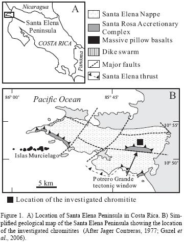
The Santa Elena Peninsula is located on the northwest Pacific coast of Costa Rica (Figure 1A). The homonymous Santa Elena Nappe (Figure 1B) consists of partially to completely serpentinized peridotites, with subordinate gabbro, thrust over the Santa Rosa accretionary complex (Tournon, 1994; Baumgartner and Denyer, 2006; Denyer et al., 2006; Gazel et al., 2006; Baumgartner et al., 2008; Denyer and Gazel, 2009). There is a general agreement to consider the rocks of the Santa Elena Peninsula as a patchwork of a dismembered ophiolite complex, probably formed in a supra–subduction zone (Denyer and Gazel, 2009 and references therein). The chromitites occur in a small area a few kilometers north of the Potrero Grande tectonic window (Figure 1B). They form irregular pods a few meters in size, associated with strongly altered peridotites covered by lateritic soil. Most of the chromitites are massive, however orbicular or leopard textures have been recognized locally. The magmatic composition of chromite shows a wide range of variation from Cr–rich to Al–rich. The Cr2O3 contents range between 43 to 67 wt% and Al2O3 varies between 6 and 23 wt%. The MgO and FeO contents range between 8–13 and 12–19 wt%, respectively, whereas the amount of Fe2O3 is negligible (less than 1.7 wt%, and in most cases 0). The TiO2 content is low, i.e. between 0.1–0.4 wt% (Zaccarini et al., in press).
3. Methodology
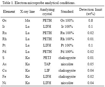
Four polished sections, with a surface area varying from 1 to 2 cm2, were prepared from each chromitite outcrop and examined by reflected light microscopy at 250–800X magnification. A total of 47 PGM have been found and all the investigated samples proved to contain at least one PGM. Their optical properties, such as color, anisotropy and reflectance were only estimated and not measured. The PGM were investigated in situ by scanning electron microscopy (SEM) and analyzed quantitatively by electron microprobe, using a Jeol JXA 8200 Superprobe, at the Eugen F. Stumpf Laboratory at the University of Leoben, Austria. The instrument operated in WDS mode, at 20 kV accelerating voltage, 10 nA beam current and with a beam diameter of less than 1 μm. The counting time on peak and backgrounds were 15 and 5 seconds, respectively. All possible peak overlaps among the selected x–ray emission lines were checked and automatically corrected, using the on–line procedure. The selected analytical conditions are listed in Table 1. Representative analyses of the PGM of Santa Elena chromitite are reported in Table 2.
Raman spectra were collected using a LABRAM (ISA Jobin Yvon) instrument at the University of Leoben. A frequency–doubled 100 mW Nd–YAG laser with an excitation wavelength of λ = 532.6 nm was used. The polarization state of the laser is consistent with the north–south direction (y–axis) of the microscope stage. Measurements were carried out with an LMPlanFI 100x/0.8 (Olympus) objective lens and they have an accuracy of 1.62 cm–1 at low Δ v (about 0 cm–1) and of 1.1 cm–1 at high Δ v (about 3000 cm–1). The following standards were employed for internal calibration: silicon (520 cm–1), polyethylene (1062 cm–1, 1128 cm–1, 1169 cm–1, 1295 cm–1, 1487 cm–1, 1439 cm–1, 2848 cm–1, 2881 cm–1), calcite (156 cm–1, 283 cm–1, 713 cm–1, 1087 cm–1, 1437 cm–1) and diamond (1332 cm–1).
4. The platinum group minerals in the Santa Elena chromitites
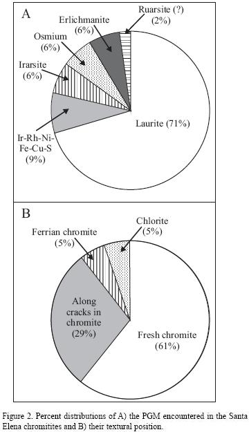
The PGM grains are always less than 10 μm in size. They consist of: 1) laurite, ideally RuS2 (33 grains); 2) unnamed PGE–Base Metals (BM) sulfides (4 grains); 3) erlichmanite, ideally OsS2 (3 grains); 4) irarsite, ideally IrAsS (3 grains); 5) osmium, ideally Os (3 grains) and 6) an unidentified RuAsS, possibly ruarsite, ideally RuAsS (1 grain). The distribution of the identified PGM is reported in Figure 2A. Regarding their textural position, the majority of the PGM were found enclosed in fresh chromite, subordinately along cracks and fissures in the chromite crystals, and a few are enclosed in ferrian chromite and in the silicate matrix composed of chlorite (Figure 2B). The PGM of the Santa Elena chromitites occur as single phase crystals or form composite aggregates in association with other PGM, base metals sulfides, clinopyroxene and chlorite.
4.1. Members of the laurite–erlichmanite series
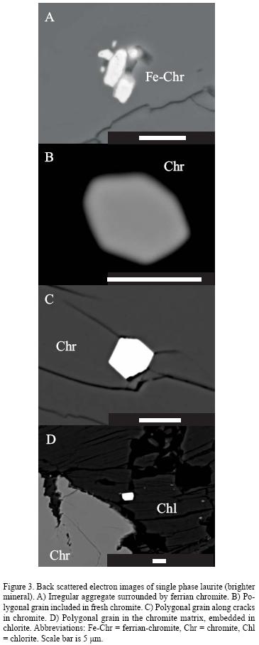
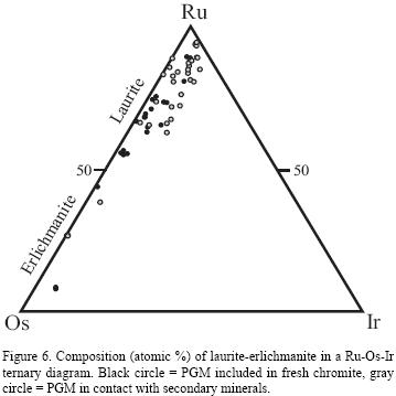
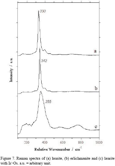
Laurite and erlichmanite are members of the RuS2 –OsS2 solid solution, with remarkable substitution of Ir for Os and Ru (Bowles et al., 1983 and reference therein). Other PGE (Rh, Pt, Pd) and base metals (Fe, Ni, Cu = BM) may occur as trace elements. Appreciable amount of As can substitute for S. Laurite is by far the most abundant PGM found in the Santa Elena chromitites. With few exceptions (Figure 3A) laurite forms polygonal grains enclosed in fresh chromite (Figure 3B), related to cracks in fractured chromite (Figure 3C). It also occurs in the altered silicate matrix in contact with chlorite (Figure 3D) or other secondary silicates. Laurite may form polyphase grains with clinopyroxene (Figure 4A), amphibole and chlorite, or with Ni sulfides (Figure 4B) and other PGM (Figure 4C). Three grains of erlichmanite were identified. One was found enclosed in fresh chromite in contact with an unknown Ir–Ni sulfide and clinopyroxene (Figures 5A and B). The other two occur as single phase grains in contact with chlorite. One of these grains is strongly zoned, showing a Ru–rich, S–poor rim (Figures 5C and D), as a result of low temperature processes. The composition of the analyzed PGM of the laurite–erlichmanite series is plotted as atomic % in a ternary diagram in Figure 6. There are no obvious differences between the PGM included in fresh chromite and those found in contact with alteration minerals (ferrian chromite and chlorite), except for a weak enrichment in Ir in the PGM associated with alteration minerals. We have obtained Raman spectra for laurite with Os>Ir (Figure 7a), erlichmanite (Figure 7b) and laurite with Ir>Os (Figure 7c). Both laurite Os>Ir and erlichmanite display well–defined absorption bands at 330 and 342 cm–1, respectively. Conversely, the laurite peak containing Ir>Os is less obviously shown in the absorption band at 355 cm–1.
4.2. Irarsite
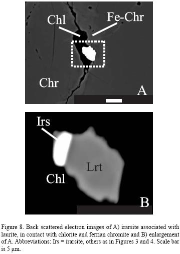
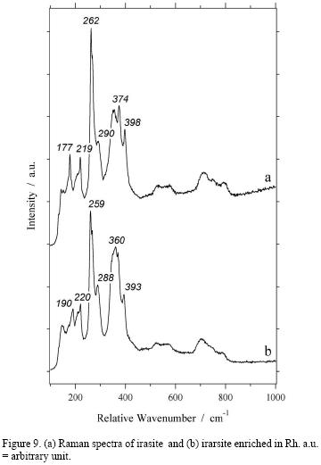
The sulfarsenide irarsite (ideally IrAsS) is the Ir end member of a complicated solid solution series that comprises ruarsite (ideally RuAsS), osarsite (ideally OsAsS), platarsite (ideally PtAsS) and hollingworthite (ideally RhAsS). Three grains of irarsite were identified, but could not be analyzed quantitatively because of the small grain size (less than 3 μm). The grains occur along cracks, in contact with chlorite, and are either single phase or composite aggregates in association with laurite (Figures 8A and B). In one case, the single–phase irarsite is characterized by a high Rh content (up to 6.9 wt%). This difference in composition is also reflected in the Raman spectra presented in Figures 9a and b. Although several absorption bands over the range of 177–398 cm–1 are clearly discernible in both analyzed irarsite grains, their intensity and shape are different.
4.3. Osmium
Osmium is one of the most common PGE alloys that occur in the podiform chromitites. It may contain appreciable amounts of Ir and Ru. In the investigated chromitites, osmium occurs as minute particles (< 1 \B5m in size) and therefore it was only identified qualitatively. Osmium occurs as small blebs attached to the external border of laurite (Figures 10A and B) or inside a polyphase grain composed of silicates (clinopyroxene and amphibole) plus Ni and Cu sulfides (Figures 10C–10F). The textural relationships support that osmium represents a low–temperature exsolution product.
4.4. Unknown PGE–BM sulfides
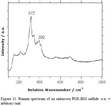
Four grains containing Ir and S as major constituents with minor, Rh, Ni, Fe, Cu were analyzed qualitatively, being only 2–3 μm in size. These PGM were encountered in fresh chromite as part of composite inclusions with laurite (Figures 4C), erlichmanite and clinopyroxene (Figures 5A and B), chalcopyrite and Ni–sulfide. In spite of a lack of quantitative analyses because of the small grain size, these PGM appear to have a characteristic Raman spectrum, as shown in Figure 11.
5. Discussion and conclusion
5.1. Magmatic conditions of PGM precipitation and their post–magmatic evolution
In ophiolitic chromitites, minerals of the laurite–erlich–manite series (RuS2 – OsS2) and Os–Ir–(Ru) alloys are the most common PGM. Most of these PGM form euhedral crystals occurring enclosed in unaltered chromite crystals. They are, therefore, classified as primary PGM, i.e. formed during the magmatic stage, prior to or concomitantly with the crystallization of chromite. According to Tredoux et al. (1995), PGE in natural magmas initially occur as a suspension of clusters of a few hundred atoms in the metallic state, without any ordered structure. Thanks to their chemical and physical properties, these clusters tend to coalesce together and adsorb particular ligands (S, As, Te, Bi, Sb) without formal chemical bonding. With decreasing temperature, the clusters form specific PGM alloys or compounds with one of the above ligands, characterized by specific crystalline structure. Subsequently, these primary PGM are mechanically entrapped by the early–precipitating minerals (i.e. chromite, olivine, pyroxene). Experimental data (Brenan and Andrews, 2001; Andrews and Brenan, 2002; Bockrath et al., 2004) supported by a great number of natural observations (Uysal et al., 2007: El Ghorfi et al., 2008 and references therein) have demonstrated that the precipitation of magmatic PGM in the ophiolitic chromitites is controlled by the following three main parameters: 1) availability of PGE in the system, 2) temperature and 3) sulfur fugacity. Sulfur fugacity increases with decreasing temperature and this variation strongly infuences the paragenesis of magmatic PGM. Therefore, at very high temperature (around 1300°C) laurite precipitates in equilibrium with Os–Ir–(Ru) alloys. Substitution of Os for Ru in laurite increases with decreasing temperature and increasing sulfur fugacity, therefore the stability field of laurite expands, to reach the composition of erlich–manite. This magmatic behavior is also registered in the compositional zoning visible in some laurite grains that generally display Os enrichment in the rim. As a consequence, the magmatic composition of minerals of the laurite–erlichmanite series can be used as an efficient tool to model conditions of PGM precipitation.
At Santa Elena, the absence of magmatic Os–Ir–(Ru) alloys and the presence of erlichmanite and abundant PGE–BM sulfides in the PGM assemblage suggest that the crystallization of PGM started at temperatures lower than 1300°C and/or relatively high sulfur fugacity.
It has been shown that magmatic PGM can be altered and modified at low temperature. In particular, minerals of the laurite–erlichmanite series can be affected by initial reduction with progressive loss of S (i.e. desulfurization process) followed, in some cases, by addition of O and Fe that allows formation of secondary Ru–alloys and Ru–Fe oxygenated compounds (Stockman and Hlava, 1984; Garuti and Zaccarini, 1997; Tsoupas and Economou–Eliopoulos, 2008; Zaccarini et al., 2009). Post magmatic alteration of laurite may also involve loss of Os and Ir, resulting in an increase of its Ru content (Zaccarini et al., 2005; El Ghorf et al., 2008).
At Santa Elena, most PGM of the laurite–erlichmanite series are characterized by similar euhedral shape and chemical composition, regardless of their textural position or association with magmatic or alteration minerals. These features indicate that the majority of these PGM were not significantly affected by metamorphism and alteration (i.e. serpentinization and lateritization). Only one grain of erlichmanite (Figures 5C and D), found in contact with chlorite in the altered matrix of the chromite, displays a Ruenriched rim. This observation suggests that this sulfide was affected by the same alteration processes, previously described by Zaccarini et al., (2005) and El Ghorf et al., (2008). Osmium occurs exclusively as small droplets inside laurite and BM sulfides (Figure 10), suggesting that it is an exsolution product, formed at low temperature.
5.2. PGM and their analytical uncertainty
It is well known that the identification of nanometer–scale minerals, such as most of the PGM associated with podiform chromitite, is a challenging target. The main reason for that resides in their size and mode of occurrence (i.e. composite polyphase aggregates) that prevent any XRD–based structural study of their crystal lattice. Optical and electron microscopy and determination of micro–hardness are the most frequently used techniques for mineralogical identification of PGM, although data in the literature show how many limitations these techniques involve. Even the most commonly used technique, electron microprobe analysis, requires accurate calibration of the instrument under the most appropriate analytical conditions (i.e. accelerating voltage, peak and backgrounds counting rates, beam diameter and beam current) in order to reduce analytical uncertainties. However, relevant limitations are expected when the grain size and mineralogical heterogeneity are of the same scale as the minimum electron beam diameter (~ 1 \B5m). In these cases, major artefacts are analytical totals much lower than the theoretical 100% and contamination from spurious fluorescence emission due to direct or secondary excitation from the neighboring minerals.
Some years ago, results were published on laser–Raman microprobe techniques that were applied for the first time to identify grains of natural PGM less than 10 microns in size from the Munni Munni layered intrusion in Australia (Mernagh and Hoatson 1995). More recently, Zaccarini et al. (2009) published Raman spectra of several PGM found in the Loma Peguera chromitites (Dominican Republic) that potentially represent new minerals. McDonald et al. (in press) obtained Raman spectrum on garutiite (Ni,Fe,Ir), a new hexagonal form of native Ni discovered in the chromitite of Loma Peguera.
Raman spectroscopy is very sensitive to the presence of covalent bonding, producing a very well–defined and visible spectrum in the material in which this type of bond is present. Garutiite is characterized by a fat Raman spectrum, thus suggesting that the possible bonds present in this new PGM are metallic or ionic.
The results published so far, although preliminarily, show that Raman spectroscopy is a fast and cost–efficient technique that can be considered an innovative and complementary methodology with a huge potential to identify and to better characterize the nanometer–scale PGM.
The Raman spectra obtained on minerals of the laurite–erlichmanite series, irarsite and unnamed PGE–BM sulfide of the Santa Elena chromitite, presented in this contribution, confirm the validity of Raman spectroscopy to distinguish the PGM. Our data suggest that this technique is also sensitive to compositional variation, particularly in the PGM characterized by a solid solution substitution such as laurite–erlichmanite series and irarsite.
Acknowledgements
Many thanks are due to R. Blanco and the staff of the National Park of Santa Rosa for their help during the field work. Many thanks also to H. Muehlhans for the sample preparation and to the University Centrum for Applied Geosciences (UCAG) for the access to the E. F. Stumpf electron microprobe laboratory. The constructive comments of Michel Dubois and Ibrahim Uysal greatly improved the quality of the manuscript. The suggestions and careful editing of Antoni Camprubí are gratefully acknowledged.
Bibliographic references
Andrews, D.R.A., Brenan, J.M., 2002, Phase–equilibrium constraints on the magmatic origin of laurite + Ru–Os–lr alloy: The Canadian Mineralogist, 40, 1705–1716.
Augé, T., Johan, Z., 1988, Comparative study of chromite deposits from Troodos, Vourinos, North Oman and New Caledonia ophiolites, in Boissonnas, J., Omenetto, P. (eds.) Mineral deposits within the European Community: Society for Geology Applied to Mineral Deposits Special Publication, 6, 267–288.
Baumgartner, P.O., Denyer, P., 2006, Evidence for Middle Cretaceuos accretion at Santa Elena Peninsula (Santa Rosa Accretionary Complex), Costa Rica: Geologica Acta, 4, 179–191.
Baumgartner, P.O., Flores, K., Bandini, A.N., Girault, F., Cruz, D., 2008, Upper Triassic to Cretaceous radiolaria from Nicaragua and northern Costa Rica – The Mesquito composite oceanic terrane: Ofoliti, 33, 1–19.
Brenan, J.M., Andrews, D.R.A., 2001, High–temperature stability of laurite and Ru–Os–Ir alloys and their role in PGE fractionation in mafic magmas: The Canadian Mineralogist, 39, 341–360.
Bowles, J.F.W., Atkin, D., Lambert, J.L.M., Deans, T., Phillips, R., 1983, The chemistry, reflectance, and cell size of the erlichmanite (OsS2)–laurite (RuS2) series: Mineralogical Magazine, 47, 465–471.
Bockrath, C., Ballhaus, C., Holzheid, A., 2004, Stabilities of laurite RuS2 and monosulfide liquid solution at magmatic temperature: Chemical Geology, 208, 265–271.
Capobianco, C.J., Drake, M.J., 1990, Partitioning of ruthenium, rhodium, and palladium between spinel and silicate melt and implications for platinum group element fractionation trends: Geochimica et Cosmochimica Acta, 54, 869–874.
Constantinides, C.C., Kingston, G.A., Fisher, P.C., 1980, The occurrence of platinum group minerals in the chromitites of the Kokkinorotsos chrome mine, Cyprus, in Panayioutou. A. (ed.), Ophiolites: Proceedings, International Ophiolite Symposium 1979: Nicosia, Cyprus, Geological Survey Department, 93–101.
Denyer, P., Baumgartner, P.O., Gazel, E., 2006, Characterization and tectonic implications of Mesozoic–Cenozoic oceanic assemblage of Costa Rica and Western Panama: Geologica Acta, 4, 219–235.
Denyer, P., Gazel, E., 2009, The Costa Rican Jurassic to Miocene oceanic complexes: origin, tectonics and relations: Journal of South American Earth Sciences, 28, 429–442. El Ghorf, M., Melcher, F., Oberthur, T., Boukhari, A.E., Maacha, L., Maddi, A., Mhaili, M., 2008, Platinum group minerals in podiform chromitites of Bou Azzer ophiolite, Anti Atlas, Central Morocco: Mineralogy and Petrology, 92, 59–80.
Elhaddad M. A., 1996, The first occurrence of platinum group minerals (PGM) in a chromite deposit in the Eastern Desert, Egypt: Mineralium Deposita, 31, 439–445.
Garuti, G., Zaccarini, F., 1997, In–situ alteration of platinum–group minerals at low temperature: evidence from serpentinized and weathered chromitite of the Vourinos complex (Greece): The Canadian Mineralogist, 35, 611–626.
Garuti, G., Zaccarini, F., Economou–Eliopoulos, M., 1999a, Paragenesis and composition of laurite from the chromitites of Othrys (Greece): implications for Os–Ru fractionation in ophiolitic upper mantle of the Balkan peninsula: Mineralium Deposita, 34, 312–319.
Garuti, G., Zaccarini, F., Moloshag, V. , Alimov, V., 1999b, Platinum–group minerals as indicators of sulfur fugacity in ophiolitic upper mantle: an example from chromitites of the Ray–Iz ultramafic complex, Polar Urals, Russia: The Canadian Mineralogist, 37, 1099–1115.
Gazel, E., Denyer, P., Baumgartner, P.O., 2006, Magmatic and geotecto–nic significance of Santa Elena Peninsula, Costa Rica: Geologica Acta, 4, 193–202.
Gervilla, F., Proenza, J.A., Frei, R., González–Jiménez, J.M., Garrido, C.J., Melgarejo, J.C., Meibom, A., Díaz–Martínez, R., Lavaut, W., 2005, Distribution of platinum–group elements and Os isotopes in chromite ores from Mayarí–Baracoa Ophiolitic Belt (eastern Cuba): Contributions to Mineralogy and Petrology, 150, 589–607.
Grammatikopoulos, T.A., Kapsiotis, A., Zaccarini, F., Tsikouras, B., Hatzipanagiotou, K., Garuti, G., 2007, Investigation of platinum–group minerals (PGM) from Pindos chromitites (Greece) using hydroseparation concentrates: Minerals Engineering, 20, 1170–1178.
Jager Contreras, G., 1977, Geologia de las mineralizaciones de cromita al Este de la Peninsula de Santa Elena, Provincia de Guanacaste, Costa Rica: San Jose, Costa Rica, University of Costa Rica, Ph.D. Thesis, 136 p.
Kapsiotis, A., Grammatikopoulos, T.A., Zaccarini, F., Tsikouras, B., Garuti, G., Hatzipanagiotou K., 2006, PGM characterization in concentrates from low grade PGE chromitites from the Vourinos ophiolite complex, northern Greece: Transactions of the Institution of Mining and Metallurgy, Section B–Applied Earth Science 115, 49–57.
Kapsiotis, A., Grammatikopoulos, T., Tsikouras, B., Hatzipanagiotou, K., Zaccarini, F., Garuti, G., 2009, Chromian spinel composition and Platinum–group element mineralogy of chromitites from the Milia area, Pindos ophiolite complex, Greece: The Canadian Mineralogist, 47, 1037–1056.
Kocks, H., Melcher, F., Meisel, T., Burgath, K.–P., 2007, Diverse contributing sources to chromitite petrogenesis in the Shebenik Ophio–litic Complex, Albania: evidence from new PGE– and Os–isotope data: Mineralogy and Petrology, 91, 139–170.
Kuipjers, E.P., Jager Contreras, G., 1979, Mineralizaciones de cromita en la Península de Santa Elena, Costa Rica: Ciencia Tecnica, 3, 99–108. Liipo, J., 1990, Platinum–group minerals from the Paleoproterozoic chromitite, Outokumpu ophiolite complex, eastern Finland: Ofoliti, 24, 217–222.
McDonald, A.M., Proenza, J.A., Zaccarini, F., Rudashevsky, N.S., Cabri, L.J., Stanley, C.J., Rudashevsky, V.N., Melgarejo, J.C., Lewis, J.F., Longo, F., Bakker, R., Garutiite, (Ni,Fe,Ir), a new hexagonal polymorph of native Ni from Loma Peguera, Dominican Republic: European Journal of Mineralogy (in press).
McElduff, B., Stumpf, E.F., 1990, Platinum–group minerals from the Troodos ophiolite, Cyprus: Mineralogy and Petrology, 42, 211–232.
Melcher, F., Grum, W., Simon, G., Thalhammer, T.V., Stumpfl, F.E., 1997, Petrogenesis of the ophiolitic giant chromite deposits of Kempirsai, Kazakhstan: a study of solid and fluid inclusions in chromite: Journal of Petrology, 38, 1419–1438.
Mernagh, T. P., Hoatson, D. M., 1995, A Laser–Raman microprobe study of platinum–group minerals from the Munni Munni layered intrusion, West Pilbara Block, Western Australia: The Canadian Mineralogist, 33, 409–417.
Proenza, J.A., Zaccarini, F., Lewis, J.F., Longo, F., Garuti, G., 2007, Chromian spinel composition and the Platinum–Group Minerals of the PGE–rich Loma Peguera chromitites, Loma Caribe peridotite, Dominican Republic: The Canadian Mineralogist, 45, 631–648.
Proenza, J.A., Zaccarini, F., Escayola, M., Cábana, C., Schalamuk, A., Garuti, G., 2008, Composition and textures of chromite and platinum–group minerals in chromitites of the western ophiolitic belt from Pampean Ranges of Córdoba, Argentina: Ore Geology Reviews, 33, 32–48.
Stockman, H.W., Hlava, P.F., 1984, Platinum–group minerals in alpine chromitites from southwestern Oregon: Economic Geology, 79, 491–508.
Tarkian, M., Naidenova, E., Zhelyaskova–Panayotova, M., 1991, Platinum–group minerals in chromitites from the Eastern Rhodope ultramafic complex, Bulgaria: Mineralogy and Petrology, 44, 73–87. Thalhammer, O.A.R., Prochaska, W., Mühlhans, H.W., 1990, Solid inclusions in chrome–spinels and platinum group element concentrations from the hochgrössen and kraubath ultramafic massifs (Austria) – Their relationships to metamorphism and serpentinization: Contributions to Mineralogy and Petrology, 105, 66–80.
Tournon, J., 1994, The Santa Elena Peninsula: an ophiolitic nappe and a sedimentary volcanic relative autochthonous: Profil, 7, 87–96.
Tredoux, M., Lindsay, N.M., Davies, G., McDonald, I., 1995, The fractionation of platinum–group elements in magmatic system, with the suggestion of a novel causal mechanism: South African Journal of Geology, 98, 157–167.
Tsoupas, G., Economou–Eliopoulos, M., 2008, High PGE contents and extremely abundant PGE–minerals hosted in chromitites from the Veria ophiolite complex, northern Greece: Ore Geology Reviews, 33, 3–19.
Uysal, I., Zaccarini, F., Garuti, G., Meisel, T., Tarkian, M., Bernhardt, H.J., Sadiklar, M.B., 2007, Ophiolitic chromitites from the Kahramanmaras area, southeastern Turkey: their platinum group elements (PGE) geochemistry, mineralogy and Os–isotope signature: Ofoliti, 32, 151–161.
Uysal, I., Zaccarini, F., Sadiklar, M.B., Tarkian, M., Thalhammer, O.A.R., Garuti, G., 2009a, The podiform chromitites in the Dagküplü and Kavak mines, Eskiseir ophiolite (NW–Turkey): Genetic implications of mineralogical and geochemical data: Geologica Acta, 7, 351–362.
Uysal, I., Tarkian, M., Sadiklar, M.B., Zaccarini, F., Meisel, T., Garuti, G., Heidrich, S., 2009b, Petrology of Al– and Cr–rich ophiolitic chromitites from the Mugla, SW Turkey: implications from composition of chromite, solid inclusions of platinum–group mineral, silicate, and base–metal mineral, and Os–isotope geochemistry: Contributions to Mineralogy and Petrology, 158, 659–674.
Vatin–Perignon, N., Amossé, J., Radelli, L., Keller, F., Castro Leyva, T., 2000, Platinum–group elements behaviour and thermochemical constraints in the ultrabasic–basic complex of the Vizcaino Peninsula, Baja California Sur, Mexico: Lithos, 53, 59–80.
Zaccarini, F., Proenza, A.J., Ortega–Gutierrez, F., Garuti, G., 2005, Platinum group minerals in ophiolitic chromitites from Tehuitzingo (Acatlan complex, southern Mexico): implications for post–magmatic modification: Mineralogy and Petrology, 84, 147–168.
Zaccarini, F., Pushkarev, E., Garuti, G., 2008, Platinum–group element mineralogy and geochemistry of chromitite of the Kluchevskoy ophiolite complex, central Urals (Russia): Ore Geology Reviews, 33, 20–30.
Zaccarini, F., Proenza, J.A., Rudashevsky, N.S., Cabri, L.J., Garuti, G., Rudashevsky, V.N., Melgarejo, J.C., Lewis, J.F., Longo, F., Bakker, R., Stanley, C.J., 2009, The Loma Peguera ophiolitic chromitite (Central Dominican republic): a source of new platinum group minerals (PGM) species: Neues Jahrbuch für Mineralogie Abhandlungen, 185, 335–349.
Zaccarini, F., Garuti, G., Preonza, J.A., Campos, L., Thalhammer, O.A.R., Aiglsperger, T., Lewis, J.. Chromite and platinum–group–elements mineralizations in the Santa Elena ophiolitic ultramafic nappe (Costa Rica): geodynamic implications: Geologica Acta (in press).
Received: 9/11/2009.
Corrections received: 13/1/2010.
Accepted: 19/1/2010.
